


Article by DENNIS HUNG
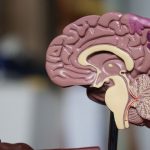
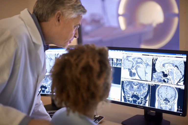
Prior to the introduction of medical imaging technology, diagnosis of internal diseases and conditions were made by surgery external examination or surgery exploration. While medicine has come an incredibly long way from using surgery to diagnose disease to the traditional 2D imaging systems that have diagnosed trillions of patients in a non invasive way. That progression prevented complications and issues that patients run the risk of having during surgeries. MRI’s and CT scans are the best they have ever been with high definition picture quality and excellent resolution that shows every detail. As helpful as that has been, at the end of the day physicians that practice in specialties involving imaging interpretation are still looking at a 2 dimensional image of a three dimensional organ or body system.
A Better Method of Surgical Preparation
In order for a surgeon to perform his or her job correctly and efficiently, they have to be able to see under the skin and possibly under other tissues that they will be operating on. This is typically done by studying the traditional 2 dimensional imaging records and at the start of surgery while the patient is under anesthesia. What 3 dimensional technology has done is made it possible for a surgeon to see the actual patients organs in a 3 dimensional image so there is no mentally piecing together the numerous angles of the 2 dimensional images. It also allows for the planning part that would usually be done at the start of the surgery after the incision is made to examine the area that is being operated on to be done prior to the surgery itself. This reduces the amount of time that the patient will be under anesthesia, which reduces the chance of complications, makes a better outcome for the patient, and reduces recovery time.
Improved Patient Comprehension and Understanding
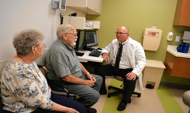
Many patients who need to make a treatment decision that often involves some kind of surgery, it is hard for them to visualize and understand the processes happening in their body that have caused them to need medical intervention. An oral explanation with a 2 dimensional picture may not be enough education for a patient to fully comprehend the severity of the disease or condition that warrants an invasive procedure. Providing the patient with the opportunity to see what the inside of their body looks like including the problem in question via 3 dimensional technology will give them more comprehensible information about the reason for the operation. The result is invaluable because the patient is the one who makes the ultimate choice about their own treatment; with this technology a more educated decision can be made by a patient in regards to their treatment options.
Quality Education Opportunities for Medical Students
3 dimensional imaging is also an invaluable tool for medical students who would otherwise need to practice on human cadavers. Because human cadavers are not readily available and because there is not a cadaver that presents with every possible disease or condition to practice treating, there is often a shortage of hands on training for unique procedures. With 3 dimensional technology in combination with a picture archiving and communication system (or PACS), a simulation and exploration of every possible surgery and disease variation can be made available for students to practice on without the difficulty of obtaining a cadaver. Medical students would get more time and opportunity to practice and learn new techniques that would have otherwise taken much longer or not been explored in a hands on approach at all.
The advancements in the medical imaging sector made possible today by 3 dimensional technology are only the beginning. The ability to put together a 2 dimensional set of images taken at different angles of the same organ to visualize what it actually looks like as a 3 dimensional object is something that the human brain is not able to do past a certain point. With 3 dimensional technology it is now possible and will continue to evolve in the coming years while improving outcomes for surgeons, patients, medical students and physicians alike.