


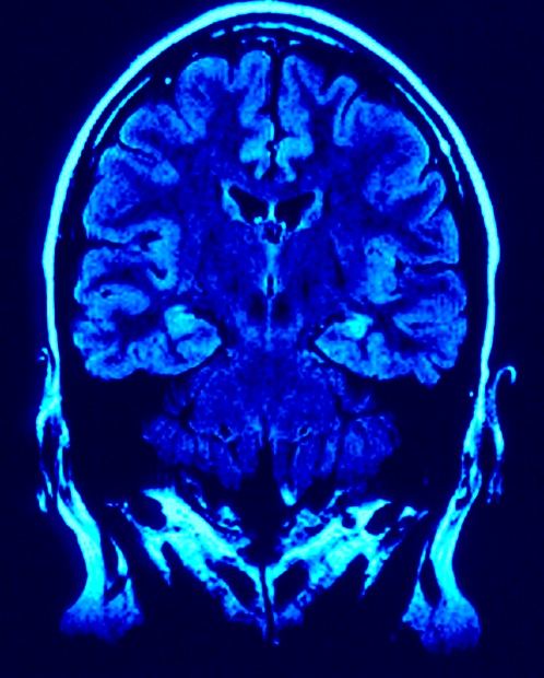
MRI, or Magnetic Resonance Imaging, is a relatively new medical technology that generates a three-dimensional image of a patient’s body. It is conveniently non-invasive and does not employ harmful radiation. More than 60 million MRI scans are performed each year.
How Do MRIs Work?
The atoms inside our bodies spin in random directions like children’s tops. An MRI employs a magnetic field twice as strong as the Earth’s to align these atoms. The field is created by an electric current flowing through a coil of wires suspended in liquid helium to reduce resistance as much as possible. This is called a superconducting magnet.
The magnetic field forces about half of the spinning atoms’ protons to point north (toward the patient’s head), and about half to point south (to his/her feet). Each northbound atom is cancelled out by a southbound atom…except for a select few. The MRI then delivers a radio frequency(RF) pulse. Remember those unmatched atoms? The RF pulse makes them spin in the other direction. When the pulse shuts off, the outlier atoms return to their original position, giving off energy that transmits a signal to a computer. The computer creates an image based on these signals.
There are three smaller, gradient magnets inside the superconducting magnet. The gradient magnets switch on and off quickly, altering the large magnetic field to zero in on a specific area of the body. The use of gradient magnets means that patients don’t have to move around inside the machine to get a precise scan.
The Improbable History of The MRI
Few people know that the entire basis of today’s MRI technology almost collapsed because of a small misunderstanding. In the early 1970s, Dr. Raymond Damadian was the first person to brave his own MRI scanning contraption, submitting his body to extreme magnetic fields in the name of science.
But…nothing happened. Damadian’s years of research, which were inspired by the Nuclear Magnetic Resonance(NMR) technology used on rocks and minerals, could’ve been for nothing. The theoretical physicists who dismissed his ideas would be proven right.
Then a graduate student with a smaller body offered to try Damadian’s scanner–and the resulting image was clear as day. The Food and Drug Administrator approved MRI technology in 1989. And that groundbreaking original machine, nicknamed the “Indomitable,” is now owned by the Smithsonian Institution.
Scientist Paul C. Lauterbur of Stony Brook University is officially credited with the invention of the MRI. Lauterbur was the first to use gradients to produce MRI images in 2D and 3D.
Why MRI?
During the 1970s, the national “war on cancer” grew urgent. Dr. Damadian’s own grandmother, who died of cancer, was a huge inspiration for his work. He estimated that tumors would emit different signals than healthy tissue, because they contained more water and thus more hydrogen atoms.
He was correct. MRI scans can detect tumors, but that’s just the beginning. They can also help doctors diagnose multiple ailments and abnormalities in the brain, spine, female pelvis, prostate, gastrointestinal tract, soft tissue, vascular system, and more.
The future of MRI is bright, as scientists look for ways to accommodate a wider range of patients and make the MRI scanning process more comfortable for all. MRI images can be used alongside other medical images like mammograms and ultrasounds to provide better patient care. PACS radiology technology allows medical organizations to efficiently store and send these diverse images together. Scientists continue to develop specialized MRI methods to obtain better results.
The Power of MRI
In 2013 alone, 108 of every 1,000 Americans had an MRI scan. That number has been on the rise since the introduction of the technology. Magnetic Resonance Imaging continues to prove its worth as one of the most elegant, painless ways of detecting health conditions before they become serious for the patient.