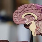


Adult mouse skeletal stem cells in the jaw revert to a more developmentally flexible state when called upon to regenerate large portions of bone and tissue, according to a study by researchers at the Stanford University School of Medicine.
The finding is the first to show that mammalian adult stem cells can march backward along the developmental timeline in a process called de-differentiation to become more primitive in response to environmental signals. In particular, the cells appeared to regress to a cell type that normally occurs within weeks of conception in humans and that give rise to the bones, cartilage and connective tissue of the head and face.
The results suggest the possibility of using naturally occurring adult stem cells, which are usually restricted to generate only a limited panel of closely related progeny, to carry out more extensive regeneration projects throughout the body — much in the way that salamanders or newts can replace entire limbs or tails.
“It’s pretty remarkable that this would happen in an adult animal,” said Michael Longaker, MD, professor of plastic and reconstructive surgery. “It changes the way we look at skeletal development and regeneration.”
A paper describing the research was published online Oct. 24 in Nature. Longaker, the Deane P. and Louise Mitchell Professor in the School of Medicine and co-director of the Stanford Institute for Stem Cell Biology and Regenerative Medicine, shares senior authorship with Howard Chang, MD, PhD, professor of dermatology and of genetics and director of Stanford’s Center for Personal Dynamic Regulomes. Graduate students Ava Carter and Ryan Ransom are the lead authors.
Aiding stunted bones
The researchers were studying a common surgical technique called distraction osteogenesis, which is often used in newborns or infants to lengthen abnormally stunted bones in the lower jaw. These malformations occur in conditions such as Pierre-Robin sequence, Treacher Collins syndrome and craniofacial microsomia.
During distraction osteogenesis, the bone is surgically fractured, and an adjustable device is inserted to gradually increase the distance between the ends of the bone over the course of weeks. This encourages new bone growth to fill in the gap and create what resembles a normally developed mandible.
“This is a very special system of regeneration that echoes what normally happens in development,” said Chang, who is the Virginia and D.K. Ludwig Professor of Cancer Genomics and a Howard Hughes Medical Institute investigator. “If you cut the bone, and stretch it, you get more bone. But this regeneration requires mechanical force. We wanted to know how skeletal stem cells respond to this kind of environmental signal.”
Recently, Stanford researchers identified the skeletal stem cell in both mice and humans. Like other stem cells that occur in adult animals, skeletal stem cells are restricted in their ability to generate different cell types. In particular, they can generate bone, cartilage and stroma (the bone’s spongy interior) to repair normal damage like fractures. But the regeneration required by distraction osteogenesis, in which the bone ends are repeatedly moved further apart, is much more extensive.
“How do they know to reform a mandible with the correct shape and function?” said Longaker. “Are they simply recapitulating normal development? And, if so, how?”
Tracking cellular reversion
Carter and Ransom used a technique developed in the Chang laboratory called ATAC-seq to identify gene switches that are turned on in mouse skeletal stem cells in response to the mechanical force of distraction. They found that the cells began to express genes normally found in cranial neural crest cells — cells that arise in humans about five to six weeks after conception and that form the bones, cartilage and connective tissue of the head and face. At the same time, the cells tamped down the expression of genes involved in normal fracture repair.
“This was really a surprise,” Longaker said. “These cells appear to revert back to a cell type responsible for forming the jaw during early development. That’s why the regenerated mandible looks like one formed in early embryogenesis.”
This is an opportunity to change how we think about the development of not just the skeleton, but also other tissues and organs.
In the absence of mechanical force to separate the bone, the skeletal stem cells repaired the fracture without expressing cranial neural crest genes.
Further research identified the focal adhesion kinase molecular pathway as a key player in the ability of the skeletal stem cells to detect and respond to mechanical force. Inhibiting this pathway abolished the ability of the cells to make new bone during distraction osteogenesis.
The finding has provocative clinical implications, the researchers believe.
“Now that we’ve identified one of the molecular pathways responsible for this developmental shift, it may be possible to target the proteins in that pathway to achieve a similar outcome without the requirement for physical force,” Carter said.
“We’re beginning to understand in detail how skeletal stem cells are likely to respond to environmental cues in humans,” Longaker said. “This is an opportunity to change how we think about the development of not just the skeleton, but also other tissues and organs. Can we go back in time after an organ is formed to trigger more extensive regeneration? This at least opens the door to that possibility.”
Longaker is a member of the Stanford Child Health Research Institute, the Stanford Cardiovascular Institute, the Stanford Cancer Institute and Stanford Bio-X. Chang is a member of the Stanford Child Health Research Institute, the Stanford Cancer Institute, the Stanford Neurosciences Institute, Stanford ChEM-H and Stanford Bio-X.
Other Stanford authors are research assistant Ankit Salhotra; former research associate Tripp Leavitt, MD; medical student Owen Marecic; CIRM scholar Michael Lopez; postdoctoral scholars Matthew P. Murphy, MD, Yuning Wei, PhD, and Ruth Ellen Jones, MD; surgical resident Clement Marshall, MD; undergraduate student Ethan Shen; assistant professor of surgery Charles K.F. Chan, PhD; and associate professor of surgery Derrik Wan, MD.
The research was supported by the National Institutes of Health (grants RO1DE026730, U24DE026914, KO8DE024269 and P50HG007735), the Howard Hughes Medical Institute, the Stanford Child Health Research Institute, the Hagey Laboratory for Pediatric Regenerative Medicine, a Steinhart/Reed Award, the Gunn-Oliver Fund and the Scleroderma Research Foundation.
Stanford’s departments of Surgery and of Dermatology also supported the work.