


The Distinct Feature Of X-Ray Technology In COVID-19 Diagnostics
In December 2019, a novel coronavirus, SARS-CoV-2, caused infection in humans in Wuhan in the Hubei province of China. The infection caused respiratory distress and is highly contagious with evidence of spreading through the air. The disease spread across continents and infection was reported from almost every country.
Imaging procedures are of great value in the COVID-19 pandemic when the question of assessment of suspected cases and determination of the course of the disease arises. While a multitude of radio diagnostic tools are employed the role of X‑ray imaging in COVID-19 infection has distinct importance.
Real-time polymerase chain reaction (RT-PCR) is the primary test for diagnosing SARS-CoV-2 infection, through running assay of throat swabs or sputum. However, since the virus’ incubation period can last up to 14 days, in the early stages, there might be a possibility of negative RT-PCR findings in the early stages of the infection. While at the onset of symptoms, computer tomography (CT) can confirm suspected cases and facilitate the prognosis of severe cases. CT has the capacity of capturing changes in the lungs of a COVID-19 patient at a high rate.
What can be the utility of X-ray imaging?
Analyzing chest X-ray findings revealed lower sensitivity for COVID-19-related lungs shadowing 25 to 69%. On the other hand, through X-Ray imaging the ability to identify the disease correctly-the specificity-was found to be up to 90%.
A significant factor in the dependability of X-ray findings could be the time elapsing between the appearance of initial symptoms and the imaging procedure. While no signs of the disease were yet noticeable in the X-ray within the initial three days after the beginning of coughing and fever, they were generally evident after 10 to 12 days. An Italian study involving 72 symptomatic patients published in mid-April 2020 appears to affirm this time factor. At the time the imaging procedure was performed, all patients were at that point under home quarantine, and came to the hospital because of worsening of symptoms. The sensitivity of the chest X-ray was reported to be 69 percent, based on the findings of the study.
Characteristic X-ray findings
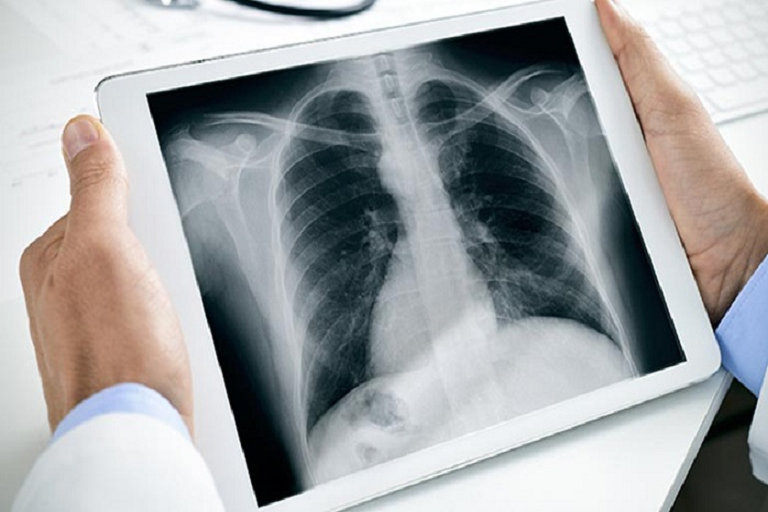
X-ray evaluation of the number of cases revealed the following characteristic X-ray findings.
Most common changes in the lungs include: – consolidation, in other words, accumulations of fluid and/or tissue in pulmonary alveoli preventing gas exchange, ground-glass opacity, and nodular shadowing.
The peripheral and lower areas of the lungs mainly gets affected.
X-ray imaging in existing circumstances
Given the availability of limited information at present, medical societies and clinical experts are still trying to frame guidelines. As far as imaging is considered, CT findings are mostly considered. It has been established that chest X-rays are insensitive in the early stages of the disease. However, if quarantined patients with advanced symptoms are examined, X-ray imaging often reveals changes in the lungs.
Considerations while making use of X-ray technology
Considering the contagious nature of the disease, contamination of the X-ray equipment and imaging machinery must be avoided. Being less complicated, X-ray systems are easier to be disinfected than CT equipment. In patients with advanced symptoms, images can be assessed in 2D for triaging COVID-19 patients. This move would reduce the burden on CT imaging techniques. Also, X-ray imaging can be performed with the patient either standing or lying down, it could be used to examine them in bed in a room specifically assigned for that purpose.
Further to this, mobile X-ray units like Kiran’s Ultisys 3.5 has the additional advantage that it can be taken directly to the patient’s bedside. This rules out the chances of viral transmission while a patient is on way to the CT room. Additionally with the integration of Picture archiving and communication system (PACS) data can be economically stored for a longer duration of time and can be transferred across longer distances thus reducing human exposure.
More info : Click here