5 Pieces of Equipment You Should Know How to Use as a Neurologist
The healthcare devices industry is one of the fastest and most lucrative industries in the world. Under this is the global neurological devices market which has been on an upward trajectory. As a neurologist, there are several devices that you need to learn how to use to execute your roles effectively. To know what devices a neurologist needs to use, you need to understand who they are.
A neurologist is a doctor who specializes in diseases of the brain, spinal cord, and peripheral nerves and muscles. Some diseases include epilepsy, stroke, Parkinson’s disease, Alzheimer’s, and multiple sclerosis. At one point or another, the neurologist may need to use a neurological device to diagnose and treat a patient.
Here are some of the pieces of equipment that a neurologist needs to know how to use.
1. Stereotaxic Frames
A stereotaxic frame is a metal device fitted over the head of a patient prepping for brain surgery. Neurologists use these frames to create a three-coordinate reference system for a precise surgical operation regardless of the patient’s movements before the procedure. These frames are often used in the surgical treatment of diseases such as epilepsy, Parkinson’s disease, and other brain-related conditions.
Stereotaxic frames are primarily used in radiosurgery to facilitate the planning phase and provide navigation during surgery.
2. TBI Impactor
A TBI Impactor is a device used for traumatic brain injury (TBI) induction. Using this device, the neurologist is able to create a neurotrauma model with a high degree of precision as to the direction of impact and the location of an injury. A TBI Impactor uses a pneumatic piston to deliver a precise injury to the patient’s neocortex to imitate the histological, physiological, and behavioral aspects of a closed head and a traumatic brain injury.
With this device, a neurologist can understand the dynamics of brain injury through a controlled impact model of the neurotrauma. Results of studies done using this device give the neurologists an in-depth understanding of disruption of the blood-brain barrier, cortical contusion, apoptosis, inflammation, hippocampal cell loss, and overall brain volume loss. This way, they are better placed to handle patients with head and brain injuries.
3. EEG Recording System
An electroencephalogram recording system is used to record detected electrical waves in the brain. This system uses electrodes to record the patient’s brain activities and transmit them to a screen for interpretation. The brain uses electrical impulses to communicate, most of which are active even when you sleep. The EEG patients and doctors see on the screen usually appear in the form of wavy lines.
As stated before, a neurologist needs to learn how to use these devices since they may come in handy when diagnosing or treating a patient. In this case, an EEG recording system is one of the most important diagnostic tests for epilepsy. It can also test for other brain disorders, including brain damage resulting from a head injury, stroke, sleep disorders, brain tumors, and inflammation of the brain.
4. Trinocular Microscopes
A neurologist’s job doesn’t end with performing surgeries or designing a treatment plan for patients; they also need to study and conduct research on the brain, spine, and nerve system to help their patients better. The Trinocular microscope is one of the best tools to help with this, especially when studying a brain tissue sample.
A Trinocular microscope is a binocular microscope with an extra slot for a camera. The camera port requires a camera adapter before you can attach a camera. Most Trinocular microscopes are designed to fit a microscopy c-mount camera. As a neurologist, you need to know how to use this device to get a clear image for a conclusive diagnosis or results.
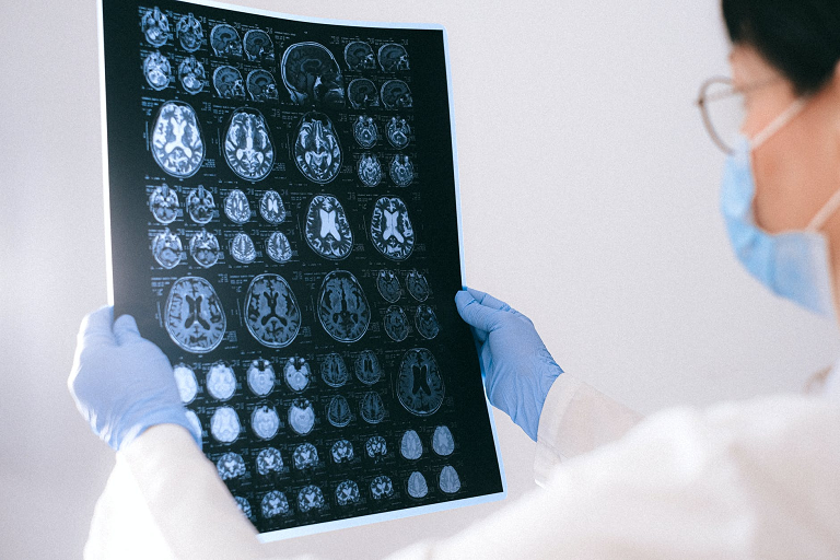
5. An MRI Scan
An MRI scan is a medical device that generates magnetic fields supported by computer-generated radio waves to create a detailed image of body organs and tissues. Doctors use this tube-like machine to examine a patient’s skeletal system, tissues, and organs. However, it is primarily used by neurologists as an imaging test for the brain and spinal cord. The device is mainly used to check for brain trauma, stroke, tumors, multiple sclerosis, and spinal cord disorders.
As a neurologist, you need to familiarize yourself with these pieces of equipment to execute your roles effectively. Most of these devices are covered under basic training in medical school, while others you may need to attend a certification program to learn.